Please activate JavaScript!
Please install Adobe Flash Player, click here for download
ePaper created 2013-08-02, 08:40:53 | version 1.25.20
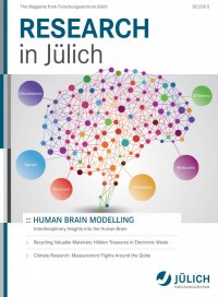
Another group of researchers at Jülich visualizes the communication pathways between neurons. Scien- tists headed by Prof. Katrin Amunts, director at the Institute of Neuroscience and Medicine (INM), have developed a method referred to as three-dimen- sional polarized light imaging (PLI) that helps them to understand the orientation of neuronal fibre tracts, the ‘hardware’ of information transmission between different brain regions. “With this three-di- mensional method of imaging the fibre tracts based on polarized light, we can produce images with a resolution in the range of thousandths of a millime- tre that are absolutely unique,” says Katrin Amunts. This network of information pathways perfectly complements another project Amunts is involved in: mapping the entire human brain. The scientist and her team have been working on a three-dimen- sional atlas of all areas of the brain for more than 15 years. For this purpose, the researchers are analysing many thousands of ultrathin tissue sec- tions per brain using state-of-the-art microscopes and image analysis techniques before reconstruct- ing the different regions of the brain in three di- mensions. Supercomputers enable scientists to deal with the huge amounts of data produced in this process. However, the digital atlas contains not only infor- mation on structural differences; it also attributes functions to different areas of the brain. Around 70% of the human brain has already been mapped. “This multimodal brain atlas can become something of a navigation system for modelling the brain,” says Amunts. It is already contributing to a better under- standing of the healthy brain, and will in future help to diagnose diseases earlier and treat them more effectively, for example. :: Prof. Paolo Carloni and his team would not have found the 5-hydroxyindole molecule as rapidly had they used a pen and paper, or experimented in the laboratory. The molecule prevents pro- teins from clumping in the brain, causing Parkin- son’s disease. “A selection procedure on a com- puter has identified this molecule from hundreds of thousands of candidates as a suitable active substance for drugs,” says the director at the In- stitute for Advanced Simulation (IAS) of Forschungszentrum Jülich. Paolo Carloni looks at processes in the brain on the smallest of lev- els: the world of molecules. For example, he ob- serves how proteins in the brain react with other substances and how taste and smell molecules take effect. The pieces in this molecular jigsaw are important for scientists in order to obtain a complete picture of our brain’s structure and the way it works. “Indeed molecules are our only weapon against neurodegenerative diseases such as Par- kinson’s and Alzheimer’s, which spread across large areas of the brain and are therefore inoper- able,” says Carloni. “The decisive factors for finding effective active substances efficiently are access to supercomputers and cooperation with laboratories to test model predictions.” :: COVER STORY | Human Brain Modelling The Power of the Tiniest of Building Blocks An Atlas with Numerous Functions Katrin Amunts, medical scientist Paolo Carloni, chemist 2|2013 Research in Jülich 9 InstituteVideo Institute